

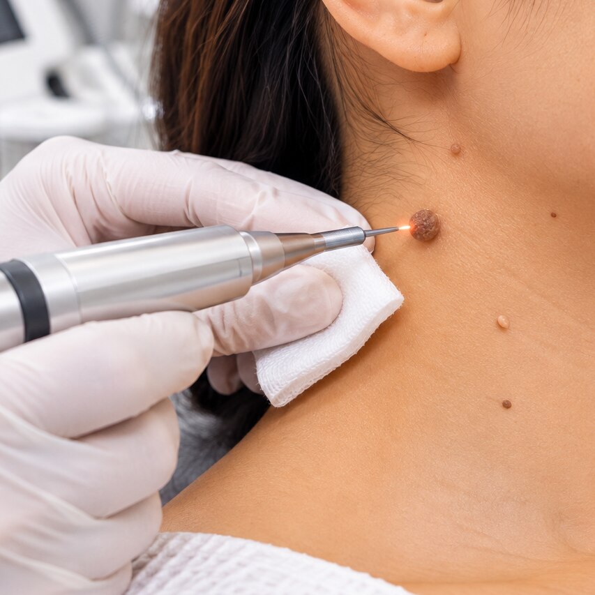
A soft tag in the neck fold, a rough wart on a finger and a dark changing mole do not carry the same implications, even when all three seem removable. By the end, you will know which signs call for examination first, when removal is optional or practical, and why the method matters as much as the decision to remove.
Key takeaways
- Do not remove a changing, bleeding, painful, or rapidly growing lesion yourself.
- A dermoscopy examination helps distinguish harmless growths from lesions needing further assessment.
- Warts need wart-specific treatment because they are caused by a viral infection.
- Ask which removal method fits the growth, its location, and your scarring risk.
First, work out what kind of growth you are looking at
Appearance can suggest the diagnosis, but it cannot reliably confirm it. A dermatologist may use dermoscopy before deciding whether removal is appropriate.
1. A skin tag, or acrochordon, is a soft, flesh-coloured or slightly darker flap hanging from a narrow stalk. It commonly appears in the neck, armpits, groin or eyelids. It is not a wart and is usually removed only if it catches, bleeds, becomes irritated or remains diagnostically uncertain.
2. A common wart, also called a verruca, feels firm and looks rough or grainy. It may show tiny black dots and interrupt the normal skin lines, unlike a smooth skin tag. Warts are caused by human papillomavirus and can spread or return after treatment.
3. Seborrhoeic keratosis usually looks like a waxy, rough or crumbly growth that seems stuck onto the skin. It ranges from pale tan to dark brown and can resemble a mole or, rarely, a skin cancer.
4. An ordinary mole is usually a stable, round or oval spot with an even colour and a fairly regular border. It may be flat or raised, but repeated catching on clothing, jewellery or a razor does not prove it is harmless.
5. Treat a new or changing pigmented lesion as suspicious until examined. Asymmetry, an irregular border, multiple colours, rapid growth, bleeding, crusting, persistent itching or a non-healing sore require assessment before removal. Do not cut, tie, burn or freeze a mole at home; this risks infection, scarring and loss of tissue needed for laboratory testing.
A changing or troublesome lesion deserves examination before removal
Removal is medically indicated when a lesion needs diagnosis, repeatedly bleeds or catches, or causes persistent pain or irritation—not simply because it looks unattractive. A clinician should establish that a benign mole, skin tag or other growth is harmless before treating it.
| Situation | What it may mean | What you should do |
|---|---|---|
| New mole | A new pigmented lesion needs assessment, especially in adulthood | Arrange a dermatology examination before removal |
| Changing mole | Growth, altered colour, bleeding, crusting or persistent itching can signal a problem | Do not treat it as a cosmetic nuisance |
| Repeated friction | A benign growth catches on clothing, jewellery or a razor and bleeds | Discuss removal after clinical examination |
| Diagnostic uncertainty | The lesion cannot be confidently identified by appearance | Ask whether sampling and histopathology are needed |
Use the ABCDE signs as a practical check: asymmetry, an irregular border, multiple colours, enlargement and evolution. Rapid growth, a non-healing sore, recurrent bleeding or pigmentation returning in a scar also warrants prompt review.
A suspicious mole should be sampled or removed in a way that preserves tissue for laboratory examination. Laser or another destructive treatment can erase that evidence. Do not tie off, cut, freeze or burn a suspected mole or skin tag at home; bleeding, infection, scarring and delayed cancer diagnosis are the risks.
Any regrowth deserves reassessment rather than repeated self-treatment.
What happens during a removal consultation
Before removal, the dermatologist must confirm what the growth is and whether destroying it would lose useful evidence. Your medical history helps: report how long it has been present, recent change, bleeding, itching, pain, previous treatments, blood-thinning medicines, allergies, poor wound healing and a tendency to form keloid scars.
1. The clinician performs a skin examination, checking the lesion’s size, shape, colour, surface, attachment and surrounding skin. They may compare it with your other moles or growths.
2. Dermoscopy magnifies structures below the surface that are not visible to the naked eye. This helps distinguish a confidently benign lesion from one needing biopsy.
3. Clinical photography can document the starting appearance, especially for a changing lesion or one being monitored before a decision. Ask how the image will be stored and compared.
4. The dermatologist explains the proposed method, expected wound care, scarring, recurrence and whether the removed tissue will go to a laboratory.
Histopathology is needed when melanoma or another tumour is possible, the diagnosis remains uncertain, or the lesion is changing, atypical, persistently bleeding or unlike your other growths.
The doctor should sample or remove it in a way that preserves tissue, then send it for microscopic examination; laser or another destructive treatment is unsuitable in that situation.
At Dr Skin N You, ask this question before treatment: “If the appearance is uncertain, will this method preserve a specimen for histopathology?” A confidently diagnosed benign tag or wart may not need laboratory testing, but the clinician makes that decision after examination.
Related service
Skin tags, warts and mole removal Skin tags, warts and mole removal Skin Tags, Warts, and Mole Removal is a safe and effective dermatological treatment offered by Dr. View service → |
How doctors choose between removal methods
Doctors match the method to the lesion’s depth, site, appearance and need for laboratory diagnosis. A benign, stalked skin tag may suit snip excision, while a superficial lesion may suit shave removal. A suspected melanoma needs preserved tissue, so diagnosis takes priority over speed or a scar-free result.
| Option | Practical difference | When it applies |
|---|---|---|
| Shave removal | Slices superficial tissue; depth can be missed. | Benign-appearing raised lesions. |
| Snip excision | Cuts a stalk at its base. | Small skin tags. |
| Punch excision | Removes a round, full-thickness specimen. | Small lesions needing sampling. |
| Elliptical excision | Removes lesion and margin; closes with stitches. | Deeper or suspicious lesions. |
| Electrosurgery | Burns tissue and seals vessels; the specimen is destroyed. | Selected benign lesions. |
| Cryotherapy | Freezes tissue; blisters and pigment change can follow. | Warts and some benign lesions. |
| Curettage | Scrapes tissue away, often followed by cautery. | Selected superficial lesions. |
| Laser treatment | Vaporises tissue, leaving little diagnostic material. | Only after diagnosis is secure. |
Laser treatment, cryotherapy, electrosurgery and curettage are unsuitable when cancer is possible because destruction prevents histopathology. A superficial shave can miss a deep melanoma, and a partial punch can miss its most informative area.
For a suspicious pigmented lesion, complete elliptical excision with a narrow clinical margin is preferred when feasible; size and site determine the final biopsy method.
Warts need a different plan from non-viral growths
A wart is an infection, not simply extra skin: human papillomavirus lives in the lesion and surrounding skin. A skin tag is a benign acrochordon, while a mole is a melanocytic growth that may need preservation for examination. Removing a wart therefore targets viral tissue; removing a tag or mole targets the growth itself.
| Growth | Usual approach | Recurrence, sites and scarring |
|---|---|---|
| Common wart | Salicylic acid or clinician-performed cryotherapy; stubborn lesions may need another method | Can recur because HPV remains nearby; face, nails, genitals and painful or resistant lesions need review |
| Skin tag | Snip removal or electrosurgery when irritated or unwanted | Usually does not recur after complete removal; neck, armpit, groin and eyelid skin can scar or change colour |
| Mole | Examination first; excision is preferred when diagnosis is uncertain | A superficial removal can allow regrowth; new pigment in a scar needs reassessment |
Home freezing products are not equivalent to medical cryotherapy. Do not apply salicylic acid or another wart product to a mole, facial or genital lesion, or any growth you cannot identify. Diabetes, poor circulation, neuropathy and immunosuppression require medical advice before treating a foot wart.
- Never tie, cut or chemically burn a suspected mole or skin tag at home; bleeding, infection and scarring are not the only risks.
- Genital warts need sexually transmitted infection care, not home treatment for hand or foot warts.
- Arrange review for wart recurrence, rapid change, persistent pain or failed treatment rather than repeatedly destroying the area.
Frequently asked questions
How can you tell whether a growth is a skin tag, wart, or mole?
Appearance can suggest the diagnosis, but it cannot confirm it. A dermatologist may examine the growth with dermoscopy before recommending removal.
When should a changing or troublesome lesion be examined before removal?
Arrange an examination before removal if the lesion changes in size, shape, or colour, or if it bleeds, hurts, itches, or becomes irritated.
What happens during a skin growth removal consultation?
The doctor asks when the growth appeared, checks its features and location, and uses dermoscopy when needed before discussing diagnosis, removal, and scarring risks.
How do doctors choose a removal method?
The choice depends on the suspected growth, its size and location, whether tissue needs testing, and the balance between effectiveness, healing time, and scarring.
Why do warts need a different treatment plan?
Warts are caused by a viral infection, so treatment focuses on clearing the wart and limiting spread rather than simply removing a non-viral growth.