

Dark spots do not all sit at the same depth or respond to the same laser setting, so successful treatment begins with identifying the pigment and its cause. By the end, you will understand how Q-switched pulses break up pigment, why wavelength and skin type matter, what a session involves, and which risks and aftercare decisions affect the result.
Key takeaways
- Q-switched pulses target melanin without continuously heating surrounding skin.
- Confirm the diagnosis before treating any changing, irregular, or unexplained dark spot.
- Ask which wavelength, settings, sessions, and aftercare your clinician recommends.
- Protect treated skin from sunlight to reduce irritation and pigmentation returning.
How a Q-switched pulse breaks up unwanted pigment
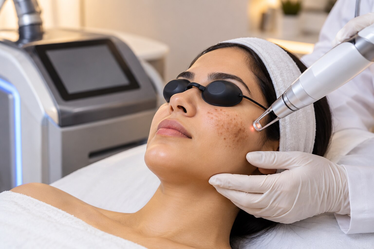
A Q-switched laser lightens a dark spot by delivering energy in nanosecond pulses, rather than heating the whole area continuously. Melanin absorbs the selected wavelength more strongly than the surrounding skin, so the pigment reaches a high temperature very quickly while nearby tissue has little time to heat.
This is the principle of selective photothermolysis: target the chromophore, confine the energy, and limit heat diffusion.
The pulse causes rapid expansion inside the pigment, producing both photothermal damage and a photoacoustic shock. That energy breaks concentrated pigment into much smaller fragments, a process described as melanin absorption and fragmentation. The body then removes the debris through immune-cell activity and normal skin turnover.
Tattoo ink follows the same basic pattern, although ink particles sit at different depths and colours respond differently to laser wavelengths.
The laser still treats the selected spot, but it does not affect every surrounding skin cell in the same way because those cells contain far less target pigment. A dark mark must be diagnosed before treatment: melanoma and pigmented skin cancers can resemble harmless spots, and laser exposure can destroy tissue needed for histopathology.
Q-switched treatment also carries risks such as blistering, crusting, infection, scarring, and temporary darker or lighter skin.
Why wavelength and diagnosis determine whether treatment is suitable
A clinician estimates pigment depth from its colour, pattern, history and dermoscopic appearance. Epidermal pigmentation versus dermal pigmentation is not always obvious by eye, so a changing, irregular or very dark spot needs medical assessment before laser; melanoma and pigmented basal-cell carcinoma can resemble harmless marks, and laser can destroy tissue needed for biopsy.
| Wavelength | Main target and depth | Key decision |
|---|---|---|
| 532 nm | Superficial epidermal melanin | Useful for freckles and solar lentigines, but blood absorbs it strongly, increasing redness, irritation and post-inflammatory pigmentation risk |
| 755 nm alexandrite | Melanin at intermediate depth | Considered for selected pigmented lesions when the diagnosis, skin type and pigment depth suit its absorption profile |
| 1064 nm Nd:YAG | Deeper dermal pigment with greater penetration | Used for deeper lesions and selected darker skin types, with careful settings to limit epidermal injury |
The choice is not a ranking from weakest to strongest. It depends on the diagnosis, skin tone, lesion depth, fluence and response. A wavelength that reaches dermal pigment can miss a superficial mark, while a highly absorbed wavelength can burn the epidermis instead of simply fragmenting pigment.
Several sessions may be needed because pigment sits at different depths and clearance must be balanced against injury. Low-fluence repeated 1064 nm Nd:YAG “laser toning” is not automatically gentle; aggressive or repeated treatment can cause mottled hypopigmentation, rebound darkening or worsening melasma. Wavelength-matched eye protection and controlled laser safety procedures are essential.
Which dark spots respond well—and which need extra caution
Freckles and solar lentigines are often good candidates because they are discrete, superficial collections of melanin. A clinician must still diagnose each dark spot first: melanoma, pigmented basal-cell carcinoma and other lesions can resemble harmless marks, and laser treatment can destroy tissue needed for histopathology.
| Condition | Suitability | Why caution matters |
|---|---|---|
| Freckles | Often suitable | They respond well when pigment is epidermal, but ultraviolet exposure can bring them back. |
| Solar lentigines | Often suitable | Treatment can target individual spots; new sun damage requires ongoing protection. |
| Post-inflammatory hyperpigmentation | Cautious | Fitzpatrick skin types IV–VI and people who mark after acne, burns or procedures face added risk of darker pigment after treatment. |
| Melasma | Highly cautious | It is a relapsing disorder driven by ultraviolet and visible light, hormones and inflammation, not isolated spots. |
Post-inflammatory hyperpigmentation calls for conservative settings, a test spot and adequate intervals between sessions. These steps reduce risk without removing it. Melasma recurrence is common after temporary laser improvement, especially when heat sensitivity, ultraviolet exposure or inflammation remains; laser alone can also worsen the condition. Topical treatment, broad-spectrum sunscreen and trigger control remain central.
Sunscreen is part of treatment, not optional aftercare. Tinted products containing iron oxides add protection against visible light, which matters particularly for melasma. Q-switched treatment will not correct redness, active acne, eczema or visible vessels; control those causes first.
What happens during treatment and in the days afterward
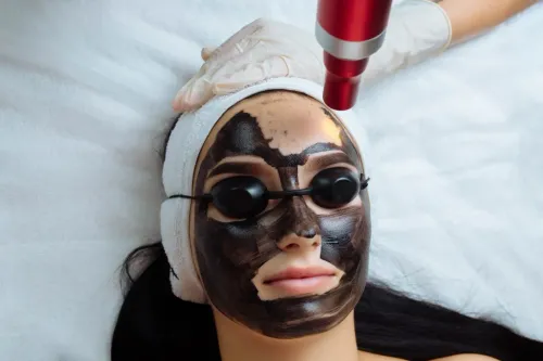
Expect a short clinical procedure: the area is examined, cleansing removes makeup and oil, and wavelength-specific eye protection is fitted for you and staff. The pulses can feel like warm rubber-band snaps, with brief stinging; cooling or a topical anaesthetic may improve comfort.
1. The clinician marks the treatment area and performs test spots when your skin type, diagnosis or prior reaction makes the response uncertain. The laser then treats selected pigment rather than the entire surrounding skin.
2. A treated spot may show immediate frosting or whitening. This temporary colour change reflects the laser’s effect on pigment and usually fades within minutes.
3. Redness and mild swelling commonly follow. They often settle over several hours, though facial areas can remain pink longer.
4. Over the next few days, darkened pinpoint areas can develop crusting and peeling. Do not pick, scrub or remove flakes early; let the surface separate naturally.
Wash gently, apply the advised bland ointment or moisturiser, and use broad-spectrum SPF 30 or higher once the skin can tolerate it. Avoid tanning, heat, exfoliating acids and retinoids until healing is complete.
Contact the clinic for increasing pain, spreading redness, pus, blisters or fever. Persistent darkening or lightening, infection, scarring and texture change are not normal recovery signs and need review, especially in Fitzpatrick skin types IV–VI or if you commonly develop dark marks after injury.
How to judge the treatment plan before you commit
Choose the plan that explains why you need a particular number of sessions, not one that sells a fixed package. The clinician should identify the diagnosis, skin type, prior reactions and pigment depth before selecting fluence, wavelength and treatment intervals.
| Plan feature | What a sound plan includes | Warning sign |
|---|---|---|
| Number of sessions | An estimate revised after each response | Guaranteed clearance in one visit |
| Treatment intervals | Enough time for redness, crusting and pigment change to settle | Weekly treatment without examining recovery |
| Safety testing | A test spot, conservative starting settings and review of prior post-inflammatory hyperpigmentation risk | Immediate full-area treatment despite dark marks after acne, burns or procedures |
| Protection | Broad-spectrum sunscreen, plus visible-light protection; tinted iron-oxide products matter for melasma | Aftercare limited to moisturiser |
Ask how the clinic will respond if the spot darkens, crusts or develops uneven colour. Do not pick crusts, and understand that temporary pepper-like darkening can take days to weeks to fade.
Eye protection matched to the laser wavelength must cover you and staff. Treatment belongs in a controlled clinical setting with documented laser-safety procedures, not an improvised room.
At Dr Skin N You, ask the clinician to record the test-spot result, settings and review date so later sessions follow your response rather than a preset schedule. Conservative settings, adequate intervals and strict photoprotection reduce post-inflammatory hyperpigmentation risk, especially in Fitzpatrick skin types IV–VI, but cannot remove it.
Related service
Q-switch lasers Q-Switch Laser Treatments are advanced dermatological procedures offered by Dr. View service → |
Frequently asked questions
How does a Q-switched laser break up unwanted pigment?
It delivers nanosecond pulses that melanin absorbs, heating pigment rapidly while limiting heat spread into nearby tissue.
Why do wavelength and diagnosis matter before treatment?
Different wavelengths target different pigment depths and colours, while an incorrect diagnosis can make laser treatment unsuitable or unsafe.
Which dark spots respond well to Q-switched laser treatment?
Some freckles, sun spots, and selected pigment conditions respond well, but changing or irregular lesions require medical assessment before cosmetic treatment.
What happens during and after Q-switched laser treatment?
You may feel brief snapping or stinging, followed by temporary redness, darkening, crusting, or sensitivity as the treated pigment clears.
How can you judge a Q-switched laser treatment plan?
Ask for the diagnosis, wavelength, fluence, expected sessions, risks for your skin type, aftercare instructions, and a plan for complications.